Financial Support for #CourageousCaitie
Fundraising campaign by
Michelle Fabella
-
US$0.00raised of $100,000.00 goal goal
No more donations are being accepted at this time. Please contact the campaign owner if you would like to discuss further funding opportunities
Faced with this diagnostic dilemma, 3 year old #courageouscaitie continues to fight and remain strong every single day in the face of multiple blood tests, biopsies and seemingly endless transfusions!!!
Rare diseases are not only difficult to diagnose, the mounting hospital bill can be very expensive as we continue to search for answers!
Together, let us pray, support and make a difference for Caitie!!! If the Lord touches your heart to give.
Thank you so much!
May God's favor be upon you and may He richly reward the goodness of your heart! ❤
" A generous person will prosper; whoever refreshes others will be refreshed." Proverbs 11:25
Update: ** They are now in Singapore- National University Hospital to undergo series of laboratory tests to further determine Caitie's medical condition. They are seeking for your financial help to help them surpass all of these. **
Let us help this brave soul. Thank you.
History:
CLINICAL ABSTRACT
Patient’s Name: Lucas, Caitlyn
Age/ Sex: 3/F
Date of admission: 2/8/16
Attending Physician: Dr. Ty Sy
CHIEF COMPLAINT: Loose Stools
HISTORY OF PRESENT ILLNESS:
2 years PTA, the patient had swelling of both feet with multiple erythematous papules over lower extremities, with no fever noted. Several unrecalled work ups were done, which revealed elevated white blood cell count. Impression at that time was cellulitis and the patient was admitted at St. Luke’s Medical Center where she was given unrecalled antibiotics.
Interval history showed intermittent appearance of erythematous papules and patches over upper and lower extremities, with some lesions becoming erythematous, hyperpigmented or violaceous over time. This was associated with swelling of bilateral ankle joints and difficulty ambulating. The patient was prescribed different unrecalled creams for the skin lesions.
1 year PTA, the patient was noted to have low hemoglobin levels and was prescribed iron supplementation of unrecalled dosage for 6 months. There was also noted persistence of skin lesions at this time.
4 months PTA, patient had prolonged cough lasting for one month. The patient was prescribed Cefuroxime of unrecalled dosage for 10 days.
3 month PTA, the patient was advised to consult a pediatric immunologist because of poor weight gain and short stature. The patient was advised to have PPD test done which showed a positive result.
2 month PTA, the patient had chest x-ray done which showed negative result. The patient was started on Isoniazid at 11.7 mg/kg/day. Patient was referred to pediatric dermatologist, rheumatologist and infectious disease specialist for the skin lesions, all of whom had a working impression of Erythema Nodosum as a reaction to Tuberculosis. Patient was started on Rifampicin at 13.3 mkday and Pyrazinamide at 25 mkday. Patient was subsequently admitted for further work-up to rule out inflammatory bowel disease. Several work-ups were done, including biopsy of nodules and bone marrow aspiration.
5 weeks prior to admission, the patient had intermittent episodes of fever, with noted appearance of new nodules, initially presenting as erythematous lesions then rapidly progressing to a purplish hue located on the face, back, and soles of feet associated with difficulty walking and occasional joint pains. Patient was admitted as a case of erythema nodosum. Ancillaries done were flat plate of the abdomen, which showed adynamic ileus and fecal stasis; CT scan of the chest and abdomen was ordered ( see results ).Because IBD was considered during this time, anti-saccharomyces antibodies and anti-nuclear antibody were requested.. Patient also underwent bone marrow aspiration biopsy and excision biopsy of the right leg. She was discharged with the following diagnosis: Reactive leucocytosis secondary to pulmonary tuberculosis; erythema nodosum; bone marrow failure probably secondary to chronic inflammatory disease.
Interval history revealed persistence of symptoms, now associated with episodes of blood-streaked, loose watery stools and blood-streaked vomiting. The patient was brought to her AMD, and gastroscopy and colonoscopy was advised to rule out inflammatory bowel disease. 4 weeks prior to admission, the patient was readmitted and referred to the following subspecialties: pediatric hematology, gastroenterologist, immunologist and geneticist for further work up and management. Impression at this time was hemophagocytic lymphohistiocytosis. She underwent right leg nodule biopsy, was started on steroids and was discharged for further workup as outpatient,
Seventeen (17) days prior to admission, the patient had low-grade fever associated with five to six episodes of loose watery stools/day and skin lesions. The patient was brought to St. Luke’s for second opinion and was admitted under the Hema-Oncology Service (Dr. Racho) with a plan to do lymph node biopsy and a repeat BMA.
Sixteen (16) days prior to admission, the patient was transfused with two (2) units of platelet conc for thrombocytopenia ,. . Peripheral blood smear showed blasts and atypical cells. Bone marrow aspiration biopsy was done. . Comprehensive leukemia panel and flow cytometry was gain requested。
Fifteen (15) days prior to admission, the patient was febrile, had loose bowel movement, and was noted to have increased abdominal girth.
Fourteen (14) days prior to admission, one unit of packed RBC was given for anemia and . Lymph node excision biopsy was done, pending results.
Thirteen (13) days prior to admission, the patient was again given one (1) unit of platelet concentrate.
Twelve (12) days prior to admission, the patient was noted to have persistent loose watery stools w/ blood-streaks, vomiting, and abdominal pain.
Eleven (11) days prior to admission, stool culture and sensitivity revealed Citrobacter freundii. Fecalysis showed pus cells 2-4, no ova and parasites, Primary Immunodeficiency Panel III revealed 13.7% lymphocytes, 18.8% monocytes, 67.5% granulocytes, normal CD3, CD4, CD8, CD4:CD8, CD16+56, CD20, and CD19 expression profiles. Serum IgG and total serum IgE were elevated (200 and 300 IU/mL, respectively), while serum IgA and Complement 3 were decreased (35.4 and 40 IU/mL, respectively). Serum IgM was within normal limits.
Ten (10) days prior to admission, the patient was noted to be pale and tachycardic. Fluid resuscitation and oxygen supplementation were done.
Nine (9) days prior to admission, there was persistent loose bowel movement which was associated with increased abdominal girth.
Eight (8) days prior to admission, the patient was referred to the GI service and the patient was given Racecadotril (Hidrasec).
Seven (7) days prior to admission, the patient was referred to the Infectious Disease Service. The patient was transfused with two (2) units of platelet concentrate. IV antibiotics was started.
Six (6) days prior to admission, the patient was noted to be febrile. Chest x-ray showed minimal streaky densities in both lung bases which may relate to subsegmental atelectasis.
Four (4) days prior to admission, the patient was referred to the Nutrition Service for persistent, uncontrolled loose bowel movement.
Three (3) days prior to admission, the patient was started on total parenteral nutrition with Aminosteril 6% and intralipid which was subsequently discontinued due to fever.
Two (2) days prior to admission, the patient was started on Somatostatin , Albumin transfusion and was 2 vial of oral IVIG was given x2days. No improvement with the LBM was noted.,. Primary Immunodeficiency Panel I revealed 9.0% lymphocytes, 21.8% monocytes, and 69.2% granulocytes. Expression of CD3, CD4, and CD8 were decreased (34.30%, 21.91%, and 10.23%, respectively. CD4:CD8 and CD16+56 expression were within normal limits. CD20 and CD19 expression was elevated at 66.87%and 65.48%, respectively.
One (1) day prior to admission, the patient decided to be readmitted at our institution.
REVIEW OF SYSTEMS:
GENERAL: see HPI
HEAD: no lacrimation, no aural discharge, no rhinorrhea, no epistaxis
RESPIRATORY: no cough, no difficulty of breathing
CARDIOVASCULAR: no cyanosis
GASTROINTESTINAL: see HPI
GENITOURINARY: no dysuria, no changes in color of urine
ENDOCRINOLOGY: no polyuria, no polydipsia, no polyphagia
NERVOUS/BEHAVIOR: no tremors, no paralysis
MUSCULOSKELETAL: see HPI
HEMATOLOGIC: see HPI
PAST MEDICAL HISTORY:
Dengue – 2014, lowest platelet count=60
PTB – 2015
HFMD – July 2015
FAMILY HISTORY
Breast CA – father’s side
Diabetes – father’s side
IMMUNIZATION HISTORY
Complete as recalled
PHYSICAL EXAMINATION ON ADMISSION
General Survey: awake, weak looking.
Anthropometrics: Wt. 11.45 kg, Ht: 98 cm
Vital Signs: BP 90/60, PR 133 bpm, RR 24 cpm, Temp 37°C
Skin: (+) hyperpigmented rash on the face, upper and lower extremity
HEENT: slightly sunken eyeballs,,pink palpebral conjunctivae, anicteric sclerae, moist buccal mucosa
Chest: symmetric chest expansion, clear breath sounds
Heart: no heaves, lifts, thrills, no murmurs.
Abdomen: globular, normoactive bowel sounds, tense, distended, fish mouth umbilicus, palpable liver edge 2cm below the subcostal margin, (+) splenomegaly
Extremities: grade 1-2 pitting edema below knee
NEUROLOGICAL EXAM
Awake, alert
CN1: was not assessed
CN2: pupils equally reactive to light
CN3, 4, 6: intact EOMs
CN5: no sensory deficit, can clench teeth
CN7: no facial asymmetry
CN8: no gross hearing deficit
CN9, 10: intact gag reflex
CN12: tongue midline
Motor: moves all extremities
Sensory: no deficit
DTRs: not assessed
No meningeal signs
Course in the ward:
Upon admission, patient was put on diet for age, and was monitored closely. Venoclysis was started and adjusted accordingly. She was referred to a pediatric intensivist and pediatric gastroenterologist for co-management. Diagnostics were ordered as follows: complete blood count with platelet (CBCP), sodium, potassium, magnesium, calcium, SGPT, CT scan of the whole abdomen and chest with triple contrast and stool culture and sensitivity. Esomeprazole was started. CBCP revealed leucocytosis (90), neutrophilic predominance (60), with leftward shift stabs (8), myelocyte (7), blast (2), juveniles (8), and thrombocytopenia (10). Patient was given platelet transfusion and albumin transfusion of which the patient tolerated well. NGT feeding with breastmilk was advised and vitamin A and folic acid were also given. Plan was to undergo colonoscopy once patient has been nutrionally built-up. During this time, patient still had loose stools, afebrile, with 8cm liver span, splenomegaly, decreased bibasal breath sounds, 97% at room air, abdomen soft, distended with fish-mouth umbilicus. Due to the blood picture, pediatric intensivist assessed the patient to have possible gastrointestinal and/or blood infection and AML in evolution. The following laboratory examinations were also requested: PT, PTT, BUN, Creatinine, Blood C/S with ARD, CRP, BUA, LDH, CBCP with PBS, iCa, TPAG. Piperacillin-tazobactam, and zinc syrup were started. Mupirocin ointment was also applied on all punctured sites. Patient was then referred to a pediatric hematologist. Laboratory examination revealed deranged PT/PTT, elevated BUN, BUA, normal creatinine, low albumin and calcium. CBCP showed leucocytsosis of 62.8 with neutrophilic predominance and leftward shift, and thrombocytopenia at 28. PBS revealed increased WBC count with predominance of segmenters. Bands, myelocytes, blast and juveniles are also noted. RBC count is normal, normocytic normochromic, normocytic hypochromic. Platelets are decreased.
On the second hospital day (2/9/16), vitamin K was started. Fresh frozen plasma was also transfused of which the patient tolerated well. CFB was noted to be at +1189; hence, furosemide was given along with Allopurinol for elevated blood uric acid. During this time, patient still had loose stools. Pediatric hematologist assessed patient to have Bone marrow failure, to consider acute leukemia. Hydration was continued accordingly and repeat comprehensive leukemia panel was suggested but was not done. CT Scan with contrast of the chest revealed almost complete resolution of the lymph nodes in the subcarinal, perihilar and prevascular areas. Several prominent axillary lymph nodes are again seen bilaterally. The lungs remain clear with no evident infiltrates or mass. The heart is not enlarged. There is no pleural of pericardial effusion. The large airways, vascular, and osseous structures are not unusual. Whole abdomen: there is significant decrease in the midhepatic length, presently measuring 8.6 (previously 11.4cm). Contour is smooth and no focal lesions are demonstrated. No dilated intrahepatic bile ducts are seen. There is no significant change in the degreee of splenomegaly. Suspicious tissue densities are again noted in the paraaortic and aortocaval regions. Gallbladder, pancreas, adrenal glands are unremarkable. Bowel cannot be optimally assessed due to lack of oral and recatal contrast. Kidneys, urinary bladder are normal. No intraabdominal mass, ascitis or mesenteric thickening seen. Vascular, osseous and the rest of the abdomen structures are not unusual. Abdominal circumference was measured every 6 hours which range of 52-53cm. Patient was stable, still with loose stools.
On the third hospital day (2/10/16), patient was again given albumin with furosemide. Esomeprazole was discontinued. Stool culture revealed no enteropathogen.
On the fourth hospital day (2/11/16), last dose of Piperacillin tazobactam was given. Platelet apheresis was suggested. Patient had persistent loose stools, IVF was ordered to be consumed and replace all loses with ORS via NGT. CBCP, PT, PTT, Na, K, TPAG, anti-dsDNA were requested. Patient was scheduled for colonoscopy once target blood count is reached. However, results of CBC showed otherwise. There was still leucocytosis with leftward shift and thrombocytopenia. During this time, the increase in white count, with anisocytosis, leftward shift, toxic granules, cystoplasmic vacuoles, immature-looking cells, atypical lymphocytes, and decreased platelets. Patient was assessed by pediatric hematologist to have possible primary immunodeficiency, probably leucocyte adhesion deficiency. IVIG and platelet apheresis were suggested.
On the fifth hospital day (2/12/16), patient received 1 unit of apheresed platelet. . Blood CS revealed negative findings. Patient was referred to rehabilitation service. She was started on rehabilitation program to prevent further deconditioning. Patient was started on range of motion exercises and gradual mobilization. Patient tolerated it well. During this time, patient was hemodynamically stable; hence, pediatric intensivist officially signed out. Albumin was again transfused.
On the sixth hospital day (2/13/16), patient had abdominal distension; hence given albumin and vitamin K one dose. However, during albumin infusion, patient developed fever at 38.1 with chills and pallor. Patient had normal blood pressure at 90/60, HR of 120, harsh breath sounds, full and equal pulses. Infusion was immediately stopped and patient was given paracetamol and diphenhydramine. Patient remained stable thereafter.
On the seventh hospital day (2/14/16), patient was given 2 vials of IVIG (1g/kg), however, patient developed transfusion reaction presenting as febrile episode at 39.1C on the first vial. Patient also complained of pain on IV site. Transfusion was immediately stopped, IV was removed and reinserted and patent was given paracetamol and monitored closely.
On the eighth hospital day (2/15/16), after 4 hours of being afebrile, patient was given another vial of IVIG of which the patient tolerated well. The patient was transfused 1 aliquot (500mL) of platelet apheresis and started on Nutramigen and protein supplementation (Aminosteril).
On the ninth hospital day (2/16/16), repeat bloodwork showed anemia with a Hgb level of 6.6, hypoalbuminemia with a serum albumin level of 17.60, and hyponatremia with a serum sodium level of 132.50. The patient was transfused 125mL pRBC and protein supplementation via Aminosteril infusion was continued.
On the 10th HD, breastmilk was discontinued and shifted to Nutramigen at 15 ccq2. Intralipid restarted and TPN continued.
Presently, patient is scheduled for colonoscopy.
WORKING DIAGNOSIS:
BONE MARROW FAILURE; TO CONSIDER MYELODYSPLASTIC SYNDROME ( with Autoimmune & Paraneoplastic Component)ACUTE MYELOGENOUS LEUKEMIA; Rule out LEUCOCYTE ADHESION DEFICIENCY
**This document may not be used for medico-legal purposes**
** They are now in Singapore- National University Hospital to undergo series of laboratory tests to further determine Caitie's medical condition. They are seeking for your financial help to help them surpass all of these. **
Let us help this brave soul. Thank you.
Organizer
- Michelle Fabella
- Campaign Owner
- Metro Manila, PH
Updates: Working Diagnosis
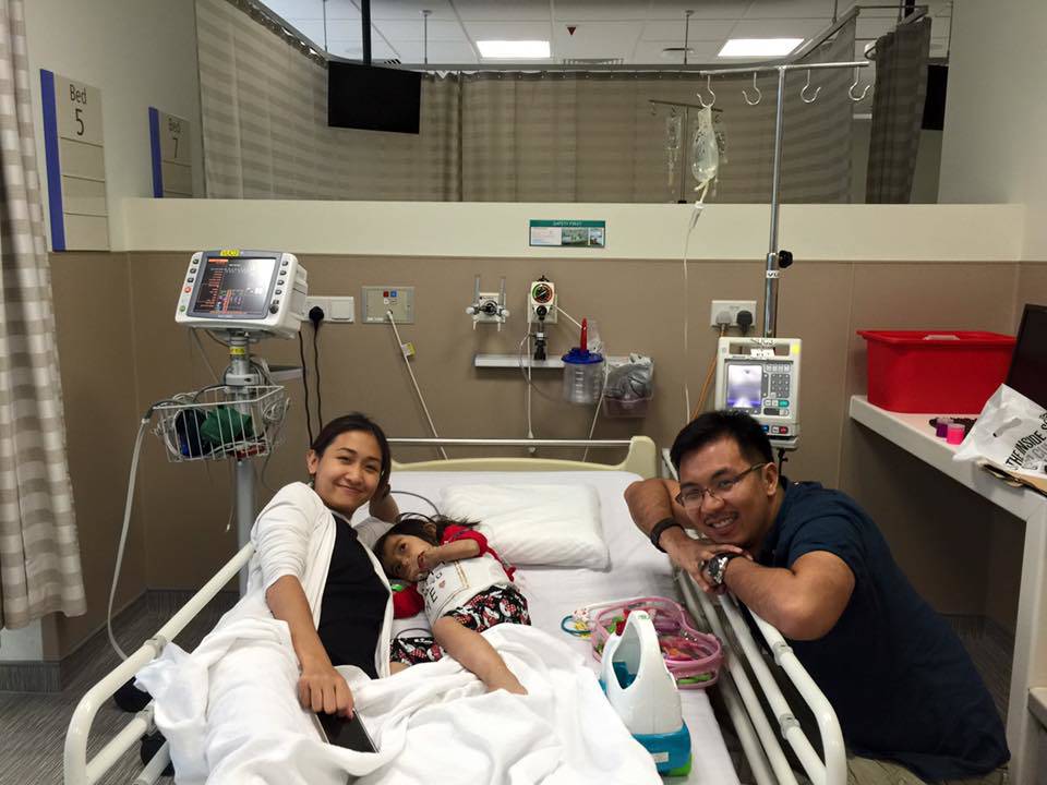
Update posted by Michelle Fabella at 07:29 amAnother platelet and blood transfusion today. She now has Singaporean blood! we are waiting for the results of all the tests which takes 4-7days running time. To finally confirm a diagnosis. We do have a working diagnosis and if it is indeed that one, we may need to do a. . . . .
National University Hospital- SG Journey
Update posted by Michelle Fabella at 06:27 amHi friends in SG! we are in NUH kentridge building ward 8b room 7. Visitors are welcome 2 at a time from 12-2pm and 5-8pm. If you are feeling under the weather or have the flu, and children under 12 years of age are not allowed to visit. Caitie's room. . . . .
Bringing God's mystery in Singapore!!
Update posted by Michelle Fabella at 06:25 amBringing God's mystery in Singapore!!“My heart rejoices in the Lord! The Lord has made me strong. Now I have an answer for my enemies; I rejoice because you rescued me. No one is holy like the Lord! There is no one besides you; there is no Rock like our God.They. . . . .

